Patient of the monthCliente del mes
Two in one week
Bladder stones are compositions of minerals of variable size in the bladder. Depending on their size and form they can stay in the bladder or migrate to the exit of the bladder. In the bladder they can cause physical irritation with inflammation, bleedings and secondary infections. When they migrate they can cause discomfort or obstruction. This latter happens especially with males because the exit canal of the bladder, the urethra, becomes narrow and inflexible at the site of the surrounding bone in the penis (see x-rays). The composition of the stones can be variable: struvite stones (most common), urate stones, oxalate stones etc. Therapeutic options depend on the size, location and composition of the stones. Stones which block the urine flow are medical emergencies and either has to be removed by retro-flush and / or operation. Stones in the bladder either have to be removed by surgery or, when they are dissolvable type of stones (struvite stones) and do not cause too much discomfort, a special diet can be fed for an extended period of time in order to try to dissolve the stones. Bladder stones are not diagnosed frequently but as a coincidence last week we had two patients with comparable urinary problems because of stone formation, which were both treated by operation, but in a different ways.
Barnie, an intelligent 10 year old male castrated Shih Tzu, who was visiting a dog kennel for only small dogs in Marbella, was diagnosed by the alert kennel owner with blood in the urine and was brought to our clinic for diagnostics and treatment. A primary and simple cystitis with males is quite rare and is normally related with prostate problems (non-castrated males) or with bladder stones. Urine testing confirmed the blood and echo-diagnostics showed bladder stones. Catheterization also revealed that the passage of the urethra was blocked just before the penis bone by some hard material. Barnie however appeared to urinate normal! An X-ray confirmed two larger bladder stones, but also showed multiple small stones (partly) blocking the urethra. With a technique called retrohydropropulsion these stones were flushed back into the bladder and all the stones were removed from the bladder by surgery and sent in for analysis. The stones appeared to be oxalate stones, which cannot be dissolved with a special diet. To prevent new stone formation it is advisable that a special diet will be fed.
A X-ray of Barnie. Two stones are located in the bladder, a few small ones are located in the upper part of the urethra.
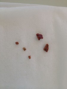
Barnie after the surgery (left). Two big stones we removed from the bladder and three little ones who were first in the urethra and after urohydropropulsion got back into the bladder (middle). X-ray after the surgery (right).
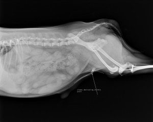
Charlie, a male non-castrated 6 year old Yorkshire terrier, was presented to our clinic 1 day earlier because he was straining when urinating. Echo-diagnostics showed an enlarged prostate, but catheterization also revealed a blockage of the urethra just before the penis bone by some hard material. An X-ray showed a small amount of small stones in the bladder and a group of stones in the urethra, lodged again just before the penis bone.
A X-ray of charlie where you can see a cluster of stones just before the penisbone.
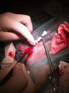
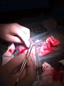
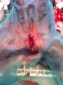
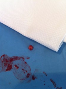
Two attempts (second attempt just before the surgery) to flush these stones back in the bladder were unsuccessful and after discussing the situation with the owner we decided to create a permanent new opening a couple of centimeters before the penis bone, an urethrostomy, which would serve as a (emergency) temporary and permanent bigger exit of the bladder, also allowing the other smaller stones in the bladder or more future stones to pass without causing obstructions. The bladder with the smaller stones was not opened, they are expected to pass the bigger exit in due time. This approach would also cause much less stress. During the surgery Charlie was castrated (also because of the large prostate) and at the site of the scrotum the new opening was created (see picture) During the surgery we were also able to grab and remove a 4 mm round stone , which was stuck firmly just before the penis bone . The other small group of stones initially present at the same location appeared to be flushed back in the bladder, which was later confirmed on the control X-ray. Charlie´s wound healed well and he is peeing well, the stones were also diagnosed as non-dissolvable oxalate stones. In a couple of weeks will be checked by X-ray whether the smaller stones have migrated outside of the bladder. A preventive diet will be fed.
On the photos you can see the operation. The third photo shows that the urethrostomy is finished and Charlie now has a new opening for urinating. ON the last picture you can see Charlie a few hours after the surgery already happy and willing to go home.
A big surprise on Valentines Day:
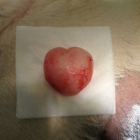
On valentines day 2013 an older blond female Labrador , Chloe , was operated on a benign , but growing , lump on the right thorax. You can imagine our and the owners surprise, when we saw the shape of the tumor at this specific day !
A nasty surprise for Dolly s family Dolly , a lovely 8 year old female mini version of a dalmation , came in for a dental the first week of November . During the check up before the pre-medication , a very large spleen was noticed and confirmed to be abnormal in shape and size by ultra-sound .The spleen is a large lymph node , positioned in the middle of the abdomen, which functions as a defense filter and as a blood reservoir . This abnormal enlarged spleens are often caused by tumors and are often detected when they start bleeding cq when the dog is in crisis. After consulting the owner , who confirmed that Dolly was not herself for some time, a blood test was performed to check 1. organ-functions and 2 . to count red-, white blood cells and blood platelets ( CBC ) This blood test revealed that her organs were alright , but that Dolly was anemic and that especially her blood platelets were extremely low . These platelets are important for the process of blood clotting , which would made it more difficult to perform a diagnostic biopsy from the enlarged spleen , because of bleeding risks. Having discussed all the options with the owner , X-rays were made from chest and abdomen and a second echo was performed to check for metastases of a possible tumor to lungs , liver and abdomen .These tests showed no visible metastases and the decision was taken to operate in order to come to a proper inspection of the abdomen and to remove the spleen , most likely the origin of the problems.
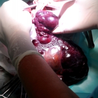
To correct the anemia , and also to add some extra blood platelets she received a blood transfusion during and after surgery. After the blood transfusion Dolly showed to be again full of life , because her batteries were recharged. The next day Dolly was operated . No visible tumor spreading s were detected in the abdomen , liver or other organs and the huge abnormal spleen was removed from the belly and sent in for pathology. Dolly went home the same evening to be with her family with special painkillers not affecting the blood clotting.The next day she came for a check-up and medication and was full of life and wagging her tail continuously . 1o days later Dolly came for stitch removal and for a blood checkup , which revealed that the anemia was resolved and that the platelets had increased to even higher than the norm .The pathology report showed that the abnormal spleen was caused by the growth of a tumor suspected to be a hemangiosarcoma, which is a malignant type of tumor of blood forming tissue . When these tumors are detected AFTER they have started bleeding , the prognosis becomes bad even on the shorter term and hopefully the early detection and removal of the abnormal spleen will keep Dolly with her loving family for many years to come !


